


Challenges today
Addressing Challenges in AI-Assisted Pathology
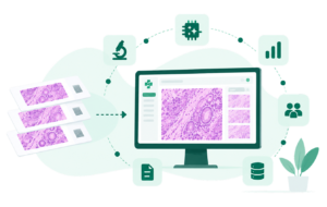
Pathologists increasingly work with complex digital images and data-intensive assessments. AI-assisted pathology can help reduce repetitive analysis, improve measurement consistency, and provide quantitative insights while keeping expert review at the center.
Managing Complex Image Analysis
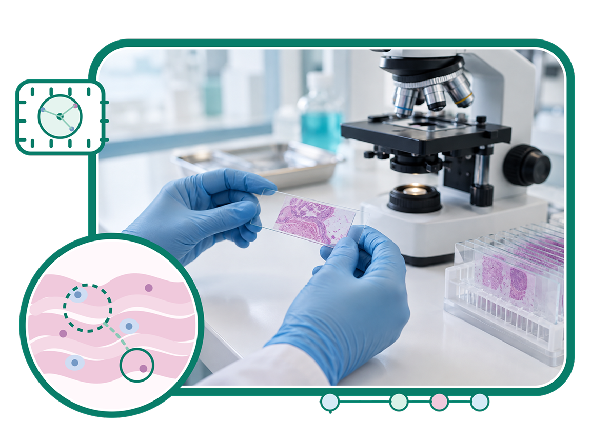
Reviewing large digital slides and identifying relevant tissue regions can require significant time and detailed manual assessment.
Standardizing AI Analysis
Manual cell counts, biomarker measurements, and image assessments can introduce variability across cases and reviewers.
Supporting Efficient Clinical Review
Pathologists need intelligent analysis tools that provide useful insights while keeping diagnostic interpretation under expert control.
Integrated AI Solutions for Pathology
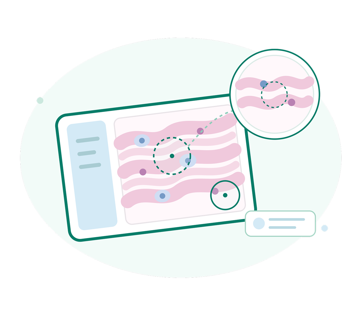
Region of Interest (ROI) Detection
Automated ROI Identification
Identify relevant tissue regions within digital slides to focus analysis on diagnostically important areas.
Guided Case Review
Help pathologists navigate large slides by highlighting regions that may require closer examination.
Efficient Image Analysis
Reduce manual searching and support faster evaluation of relevant regions across pathology cases.
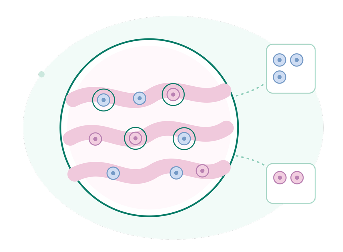
Cell Counting & Classification
Automated Cell Detection
Assist with identifying and counting cells across selected tissue regions using AI-powered image analysis.
Cell Classification
Support classification of detected cells based on defined morphological or analytical characteristics.
Consistent Measurements
Provide repeatable quantitative measurements to support standardized cellular assessment across cases.

Biomarker Scoring
Automated Biomarker Assessment
Support quantitative evaluation of biomarker expression across selected tissue regions and digital slides.
Consistent Scoring
Apply standardized image analysis approaches to support more consistent biomarker assessment.
Quantitative Insights
Generate measurable biomarker insights that can support pathology review and research workflows.
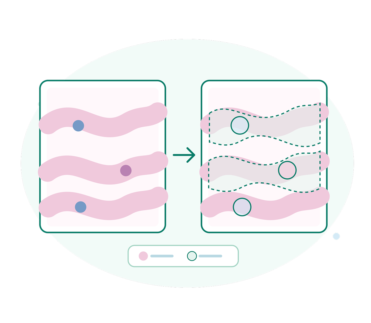
Image Segmentation
Tissue Region Segmentation
Identify and separate relevant tissue structures within digital pathology images for focused analysis.
Cellular Segmentation
Support detailed analysis by distinguishing individual cells and cellular structures within tissue regions.
Automated Image Processing
Reduce manual image preparation with AI-assisted segmentation designed for pathology analysis workflows.
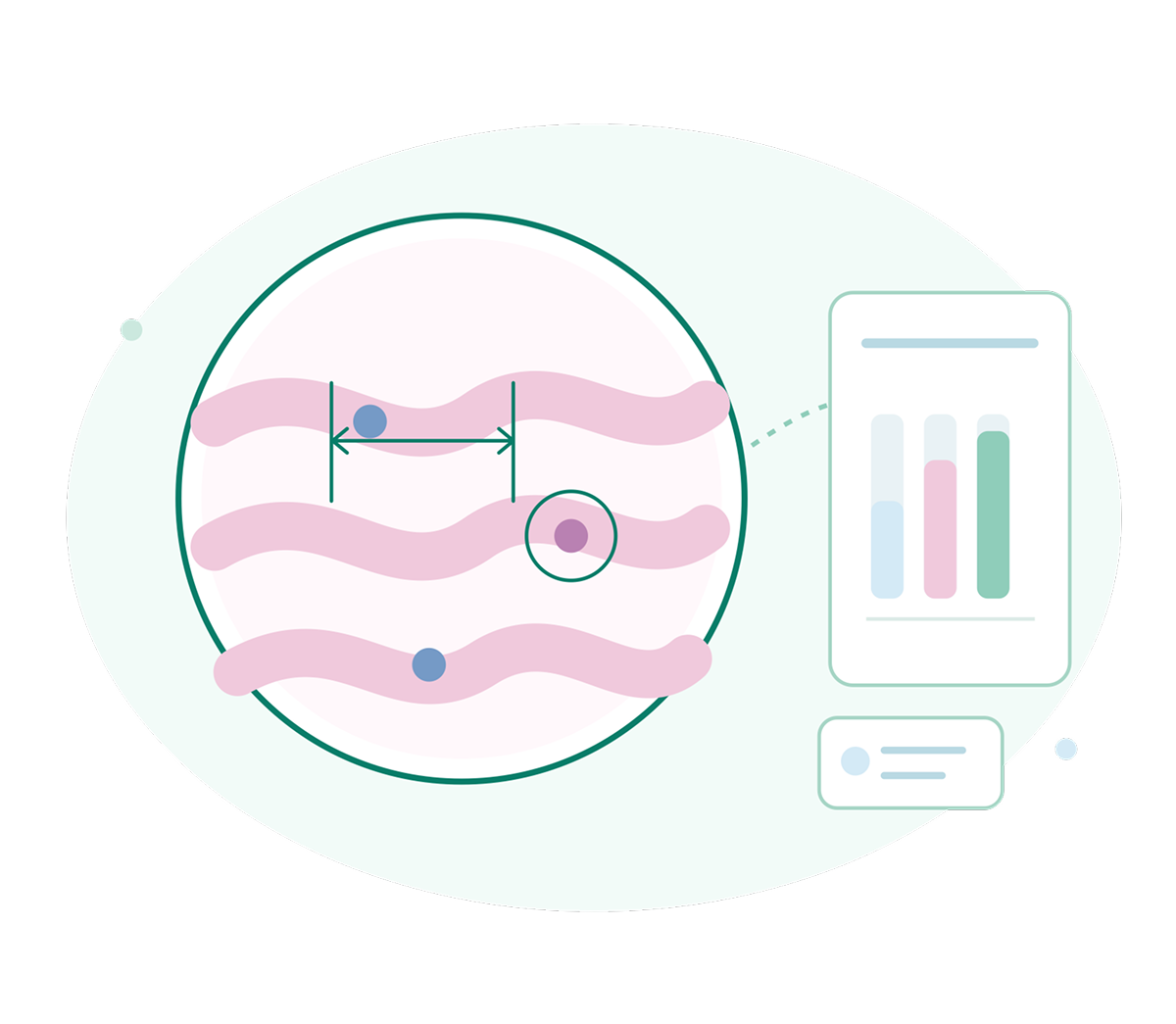
Quantitative Image Analysis
Objective Measurements
Convert visual pathology features into measurable data to support consistent image-based assessment.
Multi-Feature Analysis
Evaluate multiple cellular, tissue, and staining characteristics within defined regions of interest.
Reproducible Results
Support repeatable analysis workflows that help reduce variation in quantitative pathology assessment.
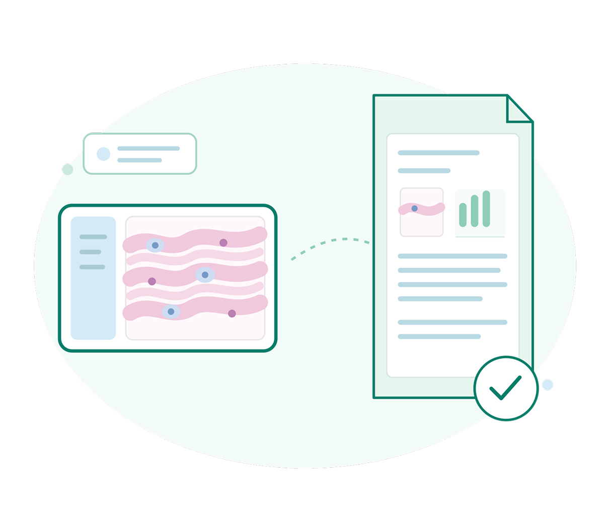
AI-Assisted Report Generation
Structured Findings
Organize analyzed pathology findings into structured information that can support report preparation.
Efficient Report Drafting
Help reduce repetitive documentation by generating AI-assisted summaries from analyzed pathology data.
Pathologist Review
Keep the pathologist in control with AI-generated content designed for expert review and finalization.
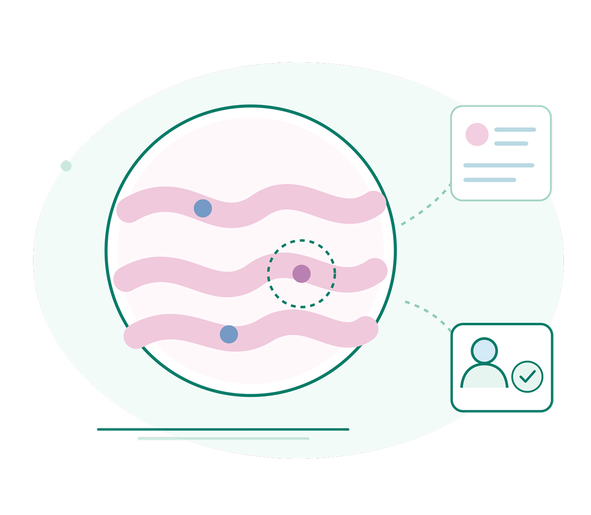
Clinical Decision Support
AI-Generated Insights
Provide AI-assisted insights from digital pathology analysis to support clinical case review.
Evidence-Based Review
Combine quantitative image findings with case information to support informed pathology assessment.
Pathologist-Led Decisions
Support clinical reasoning while keeping diagnostic interpretation and final decisions with the pathologist.