



The Rise of Virtual Spatial Proteomics in Digital Pathology
Introduction
A routine haematoxylin and eosin (H&E) slide provides pathologists with an extraordinary amount of information.
Cellular morphology, tissue architecture, stromal patterns, inflammatory distribution and the relationships between different tissue compartments all contribute to histopathological interpretation.
However, conventional H&E staining does not directly reveal the complete molecular composition of a tissue.
Many biologically important characteristics are determined by the proteins expressed by different cell populations and by how those proteins are distributed across the tissue microenvironment.
Traditionally, investigating these molecular patterns requires additional laboratory techniques, such as immunohistochemistry, immunofluorescence and spatial proteomics.
Now, a new area of computational pathology is exploring an intriguing possibility:
Can artificial intelligence estimate spatial protein expression directly from conventional H&E-stained tissue images?
This emerging research direction is often described as virtual spatial proteomics.
Using deep learning, researchers are developing models that learn relationships between tissue morphology and measured molecular profiles. These models aim to estimate how selected proteins and biological markers may be distributed across different tissue regions.
Recent research published in Nature Cancer, Nature Medicine and Cell during 2026 demonstrates how rapidly this field is progressing.
For digital pathology, this represents an important development at the intersection of whole-slide imaging, machine learning and molecular pathology.
1. What Is Spatial Proteomics?
Spatial proteomics is the study of protein expression while preserving information about where proteins are located within a tissue.
This spatial context is important because the same protein may have different biological significance depending on:
- Which cells express it
- Where those cells are located
- Whether expression is concentrated or dispersed
- How different cell populations interact
- Whether expression differs between tumour, stromal and immune compartments
Conventional protein analysis may provide information about which proteins are present and their overall abundance. However, tissue architecture and cellular location can be essential for understanding the biological significance of those findings.
Spatial proteomics attempts to preserve this information.
Depending on the technology, researchers can investigate multiple proteins across tissue regions or at cellular resolution.
This makes it possible to study complex tissue environments, including interactions between epithelial cells, stromal components and immune populations.
How is it different from immunohistochemistry?
Immunohistochemistry (IHC) uses antibodies to detect specific antigens in tissue sections. It remains an established technique in diagnostic pathology.
Spatial proteomics extends molecular investigation by enabling the analysis of multiple proteins and their spatial relationships, depending on the platform used.
The two approaches are not interchangeable. IHC remains important for established diagnostic applications, while spatial proteomics offers additional research capabilities for studying complex tissue biology.
2. What Is Virtual Spatial Proteomics?
Virtual spatial proteomics uses machine learning to estimate molecular information from conventional histopathology images.
The underlying principle is that tissue morphology contains patterns associated with cellular identity, tissue organization and biological activity.
A deep-learning model can be trained using paired datasets containing:
- H&E-stained tissue images.
- Corresponding spatial protein measurements.
- Tissue-region or cellular annotations, where available.
- Relevant biological and experimental metadata.
The model learns associations between the visual characteristics of tissue and the measured protein-expression patterns.
After training, it can generate predicted spatial protein profiles from H&E images that have not undergone the same spatial-profiling procedure.
A simplified workflow looks like this:
H&E slide → Digital image acquisition → AI-based tissue analysis → Predicted spatial protein profiles → Research and validation
The important distinction is that the AI-generated profile is an estimate, not a direct laboratory measurement.
Virtual spatial proteomics therefore does not replace the underlying experimental technology. It explores whether computational predictions can extend the research value of routinely available histology images.
3. Recent Research: AI Is Learning to Infer Molecular Information From Histology
Several recent studies demonstrate how this research area is evolving.
A. Hist2Prot: Predicting Single-Cell Spatial Proteomics From H&E
One of the newest developments appeared in Nature Cancer on 13 November 2026.
The study introduced Hist2Prot, a deep-learning framework designed to infer single-cell spatial proteomic profiles from routine H&E histology.
The researchers trained the framework using approximately 51 million paired cells from a same-section spatial proteomics dataset.
They evaluated it using independent same-platform cohorts and external cross-platform datasets, including approximately 72 million additional cells.
The study also investigated the preservation of cellular phenotypes and spatial organization, with evaluations across seven independent patient cohorts comprising 1,774 patients.
The researchers reported that Hist2Prot-derived virtual protein profiles could support molecular biomarker, prognostic and immunotherapy-response prediction tasks.
Why it matters:
This work explores whether routine histology images can provide a computational route to estimating molecular information at a much finer spatial scale.
It also highlights the importance of validation across independent datasets and different measurement platforms.
Research paper:
B. HEX: Virtual Spatial Proteomics From Histopathology
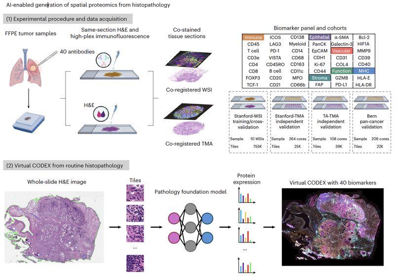
A related study published in Nature Medicine on 5 January 2026 introduced HEX, an AI model designed to computationally generate spatial proteomics profiles from standard histopathology slides.
The researchers trained and validated the model using approximately 819,000 histopathology image tiles with matched protein-expression measurements from 382 tumour samples.
The model was evaluated on 40 biomarkers representing immune, structural and functional biological programmes.
The research investigated whether spatial protein information could be estimated from conventional H&E images, offering a potential computational approach to biomarker discovery.
Why it matters:
HEX demonstrates how AI-based image analysis may help researchers investigate protein-expression patterns without requiring every analysed slide to undergo the same extensive spatial-profiling process.
However, predicted expression must still be assessed against appropriate experimental measurements.
Research paper:
C. Path2Space: Predicting Spatial Gene Expression From H&E
Another important development appeared in Cell on 8 May 2026.
The study introduced Path2Space, a deep-learning model designed to predict spatial gene expression directly from H&E-stained histopathology images.
Unlike spatial proteomics, which investigates proteins, spatial transcriptomics measures gene-expression patterns while retaining their location within tissue.
Path2Space was developed to predict the spatial expression of thousands of genes from routine histopathology images.
The researchers explored how the predicted spatial profiles could be used to identify tissue subgroups and investigate associations with survival and treatment response.
Why it matters:
This research demonstrates how AI-based tissue analysis is expanding beyond conventional morphological classification toward the computational estimation of molecular information.
It is important to distinguish predicted gene expression from directly measured transcriptomic data. The two should not be treated as equivalent.
Research paper:
AI-predicted spatial transcriptomics unlocks breast cancer biomarkers from pathology — Cell (2026)
D. OmiCLIP: Connecting Histology With Transcriptomics
Published in Nature Methods in May 2025, OmiCLIP introduced a visual–omics foundation model designed to connect H&E images with transcriptomic information.
The research used approximately 2.2 million paired tissue-image and transcriptomic patches from 1,007 samples across 32 organs.
The model was developed to learn relationships between tissue morphology and gene-expression information.
The associated Loki platform explored applications including tissue alignment, annotation, cell-type decomposition, image–transcriptomics retrieval and gene-expression prediction.
Why it matters:
OmiCLIP illustrates how foundation models can learn relationships between visual tissue patterns and molecular information across multiple tissue types.
It also demonstrates the importance of large, carefully paired datasets when developing models for computational pathology.
Research paper:
4. Why Does the Tissue Microenvironment Matter?
A tissue section is not simply a collection of individual cells.
It is an organized biological environment in which different cell populations interact with one another.
For example, the tissue microenvironment may contain:
- Epithelial or tumour cells
- Immune-cell populations
- Fibroblasts and other stromal cells
- Blood vessels
- Extracellular matrix
- Inflammatory regions
- Areas of necrosis or tissue degeneration
The distribution and interaction of these components can influence biological behaviour.
Two tissue regions may appear morphologically similar while showing differences in their molecular composition.
Similarly, two specimens with comparable overall biomarker expression may have different spatial distributions of the relevant proteins.
This is one reason spatial analysis is attracting attention.
Instead of asking only whether a marker is present, researchers can investigate:
- Which cells express the marker?
- Where is the expression concentrated?
- Are particular cell populations located near one another?
- Does expression differ between tissue compartments?
- Are there spatial patterns associated with particular biological characteristics?
AI-based spatial profiling could help researchers investigate these questions across larger collections of digitized tissue images.
5. Potential Applications in Computational Pathology
Virtual spatial proteomics is still an evolving research area. Nevertheless, several potential applications are attracting interest.
A. Biomarker discovery
Researchers may use predicted spatial profiles to identify candidate biomarkers and investigate their distribution across tissue regions.
This could help prioritize targets for further experimental investigation.
B. Understanding cellular interactions
The spatial organization of immune, stromal and other cellular populations may provide information about tissue biology.
Computational models could help identify patterns that warrant further study using direct molecular measurements.
C. Research into treatment response
Spatial molecular patterns may be associated with biological differences between patient groups.
AI-generated profiles could be investigated as research tools for identifying candidate response-associated biomarkers.
These predictions should not be interpreted as validated treatment-selection tests without appropriate clinical evidence.
D. Large-scale retrospective research
Many pathology laboratories and research institutions have accumulated substantial archives of digitized tissue images.
Computational approaches may allow researchers to investigate selected molecular patterns across archived image collections, subject to suitable data, validation and governance requirements.
E. Integrating morphology with molecular research
Virtual spatial profiling could complement conventional histopathology, IHC, molecular testing and spatial-omics research.
The objective is not to eliminate laboratory testing, but to explore how computational analysis can help connect different types of biological information.
6. The Technical Challenges Behind Virtual Spatial Proteomics
Despite the progress, translating these models into reliable research or clinical applications remains challenging.
1. Training-data quality
Models depend on appropriately paired histology images and spatial protein measurements.
Errors in image registration, tissue alignment or cellular segmentation can affect the quality of the training data.
2. Differences between laboratory environments
Staining protocols, tissue processing, fixation, scanners and image acquisition conditions can introduce variation.
A model trained using one set of laboratory conditions may not perform equally well in another.
3. Spatial resolution
Different spatial-profiling platforms operate at different resolutions.
A model trained using region-level measurements should not automatically be assumed to provide accurate single-cell predictions.
4. Biological complexity
Protein expression is influenced by many biological processes.
Morphological appearance may contain useful predictive information, but it cannot necessarily reveal every molecular characteristic of a cell or tissue region.
5. Model generalization
Independent validation across tissue types, institutions, platforms and patient cohorts is essential.
Performance on the original training dataset is not sufficient to establish broad applicability.
6. Prediction versus measurement
Perhaps the most important limitation is the distinction between an AI-generated estimate and a directly measured molecular result.
Predicted protein-expression profiles should not be presented as equivalent to IHC, immunofluorescence or validated spatial-proteomics measurements.
For diagnostic applications, appropriate analytical and clinical validation would be required, alongside regulatory and institutional assessment.
7. What This Means for Digital Pathology Platforms
Developments in virtual spatial proteomics highlight an important point: digital pathology platforms are becoming increasingly relevant to computational research.
A modern digital pathology environment needs to support more than image viewing.
It must provide a structured foundation for managing high-resolution images, organizing cases, integrating analytical tools and maintaining traceability.
Several capabilities become particularly important:
Whole-slide image management
High-resolution image storage, retrieval, visualization and organization are essential for large-scale computational analysis.
AI integration
Platforms need appropriate mechanisms to connect image-analysis tools and AI models with relevant digital pathology workflows.
Region-of-interest management
The ability to identify, annotate and revisit selected tissue regions can support image analysis and research workflows.
Structured case management
Organized case information and associated diagnostic documentation help maintain context across the workflow.
Interoperability
Connections with laboratory and hospital information systems can support the appropriate exchange of relevant data.
Security and traceability
Access controls, audit records and secure data handling are important when working with sensitive clinical and research information.
These capabilities provide the foundation for integrating current and future computational pathology technologies.
At Onward Assist, our focus is on building connected digital pathology workflows that bring together image management, case coordination, reporting and AI-assisted capabilities.
As computational pathology evolves, platforms that can support the responsible integration of new analytical technologies will become increasingly important.
8. The Future: From Morphological Patterns to Spatial Molecular Intelligence
The development of virtual spatial proteomics represents an exciting direction in computational pathology.
Research is increasingly exploring whether information contained in conventional histology images can be used to estimate molecular characteristics that traditionally require specialized laboratory techniques.
The recent development of Hist2Prot, HEX and Path2Space demonstrates that this is no longer just a theoretical research concept.
However, the scientific challenge remains substantial.
AI-generated molecular profiles must be tested against direct measurements, evaluated across independent cohorts and assessed for their reliability in different tissue and laboratory environments.
For pathologists, these technologies may eventually offer additional ways to investigate tissue biology, identify research patterns and connect morphology with molecular information.
For digital pathology platforms, the opportunity lies in providing the infrastructure that allows images, analytical tools, case information and reporting workflows to operate together.
The next stage of digital pathology may not be limited to identifying what a tissue looks like. It may increasingly explore what its spatial molecular organization can reveal.
References and Further Reading
- The deep learning framework Hist2Prot infers single-cell spatial proteomics from histology to predict biomarkers and clinical outcomes. Nature Cancer, 2026.
https://www.nature.com/articles/s43018-026-01270-8 - AI-enabled virtual spatial proteomics from histopathology for interpretable biomarker discovery in lung cancer. Nature Medicine, 2026.
https://www.nature.com/articles/s41591-025-04060-4 - AI-predicted spatial transcriptomics unlocks breast cancer biomarkers from pathology. Cell, 2026.
https://doi.org/10.1016/j.cell.2026.04.023 - A visual–omics foundation model to bridge histopathology with spatial transcriptomics. Nature Methods, 2025.
https://www.nature.com/articles/s41592-025-02707-1 - STPath: a generative foundation model for integrating spatial transcriptomics and whole-slide images. npj Digital Medicine, 2025.
https://www.nature.com/articles/s41746-025-02020-3
Editorial note: This article discusses emerging research in computational pathology. AI-predicted molecular profiles are research estimates and should not be treated as direct laboratory measurements or validated diagnostic results. Clinical use requires appropriate analytical and clinical validation, regulatory assessment where applicable, institutional governance and professional oversight.