



A small biopsy can present a disproportionately large diagnostic challenge.
A few tissue fragments may need to support morphological assessment, immunohistochemistry (IHC), special stains, molecular testing, and additional ancillary investigations. Each test may contribute valuable information, but the available tissue is finite. Every additional section, stain, or assay must be considered in the context of the diagnostic question and the material that remains.
For pathologists, the challenge is not simply obtaining a diagnosis from limited tissue. It is preserving enough diagnostic material to answer the most important questions reliably, while minimizing unnecessary tissue consumption and avoiding preventable testing failures.
This becomes particularly important when minimally invasive sampling methods provide limited material for both initial diagnosis and subsequent biomarker assessment.
Effective tissue management requires coordinated decisions across specimen collection, gross examination, histological evaluation, ancillary testing, and communication between the pathology laboratory and clinical team.
1. Why Small Biopsies Require a Different Diagnostic Strategy
Larger specimens often provide more tissue for assessing architecture, sampling heterogeneous areas, and selecting representative blocks. Small biopsies offer fewer opportunities to resolve uncertainty through additional sections or alternative tissue sampling.
The limitations extend beyond tissue quantity.
A small specimen may contain only a few representative lesional cells, substantial inflammatory or stromal components, areas of necrosis, crush artifact, or tissue that has been compromised during collection and processing.
These factors can affect both morphological interpretation and the suitability of the specimen for downstream testing.
Three challenges commonly intersect:
- Diagnostic adequacy: Is there sufficient representative tissue to establish the nature of the lesion?
- Ancillary testing adequacy: Is enough suitable material available for the tests needed to resolve the differential diagnosis?
- Predictive testing adequacy: Can the specimen support any additional biomarker analyses required for the intended clinical application?
These questions are related but distinct. A specimen may be sufficient for an initial morphological diagnosis yet inadequate for a particular molecular assay.
The diagnostic strategy should therefore consider the complete testing pathway rather than evaluating tissue adequacy solely by whether a diagnosis can be made on the initial H&E section.
2. Begin With Specimen Adequacy and Morphological Assessment
The first step in managing limited tissue is determining what the specimen actually contains.
An initial assessment should establish whether the lesional population is represented, whether the tissue is adequately preserved, and whether the observed material is sufficient to address the clinical question.
Relevant considerations include:
- The amount and distribution of lesional tissue.
- The proportion of lesional cells relative to background cells.
- Tissue preservation and the presence of artifacts.
- Necrosis, fibrosis, inflammation, or other factors that may limit interpretation.
- Whether the sampled material represents the suspected process.
- The potential need for additional ancillary investigations.
The number of tissue fragments alone is not a reliable measure of diagnostic adequacy. A small fragment containing representative lesional tissue may be more informative than a larger specimen dominated by non-lesional components.
Likewise, cellularity must be considered in relation to the intended test. Requirements for immunohistochemistry, fluorescence in situ hybridization, and molecular assays can differ substantially.
A specimen should therefore be assessed according to both its diagnostic content and its potential to support the investigations that may be needed.
3. Tissue Stewardship: Every Section Has a Purpose
Tissue stewardship refers to the deliberate management of limited pathological material to maximize its diagnostic value.
In practice, this begins before ancillary testing is ordered. Unnecessary serial sections, broad exploratory panels, and poorly coordinated testing can consume tissue without meaningfully improving diagnostic certainty.
The objective is not to minimize testing at all costs. It is to select the investigations most likely to answer the relevant diagnostic questions while preserving material for essential downstream studies.
Use a hypothesis-driven approach
Before ordering additional stains, the pathologist should identify the leading differential diagnoses and determine what information would distinguish them.
Each test should have a defined purpose.
For example, a marker may be selected to establish lineage, distinguish between morphologically similar entities, or clarify a particular feature of differentiation. If its result would not materially change the interpretation or subsequent testing strategy, its immediate value may be limited.
Coordinate testing early
Where molecular analysis or predictive biomarkers may be required, the anticipated testing needs should be considered early in the diagnostic process.
This is particularly important when the initial diagnosis and subsequent characterization depend on the same limited tissue block.
A coordinated approach can help avoid unnecessary depletion of the most representative lesional material before the complete testing plan is established.
Preserve representative material
Tissue allocation should prioritize material that best represents the lesion and is appropriate for the intended assay.
Where relevant, the pathologist may need to consider tumor or lesional cellularity, necrosis, inflammatory background, and the distribution of the target population.
These decisions are assay-dependent and should follow validated laboratory procedures.
The guiding principle is straightforward: every additional test should contribute meaningful diagnostic information or address a clearly defined clinical requirement.
4. Immunohistochemistry on Limited Tissue: Diagnostic Value Versus Tissue Consumption
Immunohistochemistry can substantially narrow a differential diagnosis, but extensive panels may be problematic when only a small number of lesional cells are available.
A large panel may consume multiple levels of a block, leaving insufficient material for subsequent testing. This can be particularly consequential when the final diagnosis requires molecular characterization or a biomarker assay with specific tissue requirements.
A more efficient approach begins with morphology and uses markers strategically.
Pathologists should consider:
Diagnostic necessity: Which markers are essential to distinguish the leading possibilities?
Expected interpretive value: Will the result meaningfully support, exclude, or refine a diagnostic hypothesis?
Tissue requirements: How much material will be consumed, and what important tests may still be needed?
Technical suitability: Is the specimen appropriate for the proposed antibody and its validated interpretation?
Future testing needs: Could the remaining tissue be required for molecular studies or other ancillary investigations?
An unexpected or inconclusive immunophenotype should prompt reassessment rather than automatically trigger a broad expansion of the panel.
The next step may involve reviewing the H&E section, checking internal controls, selecting a more discriminating marker, or considering a complementary method.
The optimal strategy is not the largest possible panel. It is the smallest sufficiently informative panel that addresses the diagnostic problem while respecting the limitations of the specimen.
5. Molecular Testing: Why Tissue Quantity Is Only Part of the Equation
Molecular testing introduces another layer of complexity because assay suitability depends on more than the total amount of tissue available.
DNA and RNA quality, lesional cellularity, nucleic acid yield, fixation, decalcification, necrosis, and the assay’s analytical requirements can all influence whether a specimen produces a reliable result.
A tissue block may appear adequate on routine histology but still fail to meet the requirements of a particular molecular test.
Assess the specimen before testing
Where appropriate, the pathologist should identify the most representative material and assess whether the lesional population is sufficient for the intended assay.
Depending on the test, this may involve reviewing the relevant H&E section, estimating lesional cellularity, selecting a suitable region, and marking the area for macrodissection.
The requirements differ between assays, so a universal cellularity threshold should not be assumed.
Understand pre-analytical limitations
Pre-analytical variables can affect the quality of downstream molecular results.
Specimen collection, transport, fixation, processing, and storage should follow procedures appropriate to the specimen type and assay. Deviations may compromise nucleic acid quality or introduce uncertainty into the interpretation.
When a result is unsuccessful or unexpected, the cause should be investigated rather than attributing the problem automatically to insufficient tissue quantity.
Plan for sequential testing
Some cases require multiple investigations, and the order in which they are performed can influence what material remains.
Early coordination between histopathology and molecular laboratory teams can help establish a rational testing sequence and identify alternative specimen sources where appropriate.
These principles are consistent with published recommendations on optimizing small biopsy and cytology specimens for ancillary studies, including the College of American Pathologists’ guidance on thoracic small biopsy and cytology specimen handling.
6. Specimen Triage Starts Before the Tissue Reaches the Microscope
Tissue stewardship cannot be achieved by the reporting pathologist alone. It depends on the complete specimen pathway, beginning with collection and continuing through processing and testing.
Pre-analytical decisions can determine whether the specimen will support the investigations required later.
Important considerations include:
- Communication of the suspected diagnosis and anticipated ancillary testing needs.
- Appropriate specimen collection and handling.
- Use of suitable transport media and fixatives where indicated.
- Accurate specimen identification and tracking.
- Timely processing under validated conditions.
- Coordination of material for histology, cytology, microbiology, flow cytometry, or molecular testing when indicated.
The appropriate approach depends on the specimen type and clinical indication. For example, material intended for flow cytometry may require different handling from tissue intended for routine formalin-fixed histology.
When multiple diagnostic methods are anticipated, early communication can help ensure that the specimen is allocated appropriately rather than placing the entire diagnostic burden on a single tissue block.
The laboratory should establish clear protocols for situations in which limited material must support competing diagnostic requirements.
7. When the Specimen Is Insufficient: What Should Happen Next?
An insufficient specimen does not always mean that the diagnostic process must stop. However, the next step should be determined by the nature of the limitation and the clinical question.
A systematic review should establish whether the problem relates to inadequate lesional representation, poor preservation, insufficient cellularity, technical assay failure, or a mismatch between the specimen and the test requirements.
Depending on the circumstances, appropriate options may include:
- Reviewing deeper sections or existing slides where useful.
- Reassessing the most representative block or tissue region.
- Selecting an alternative validated assay.
- Evaluating suitable cytology preparations or other available specimens.
- Consulting the relevant laboratory team about testing requirements.
- Discussing repeat sampling with the clinical team when clinically appropriate.
Repeat sampling should not be recommended automatically. Its potential value must be weighed against clinical feasibility, procedural risks, and whether additional material is likely to resolve the diagnostic uncertainty.
Equally, a negative or unsuccessful molecular result should not be interpreted as evidence that the relevant molecular alteration is absent when the assay has not produced an informative result.
Clear reporting should distinguish a genuinely negative result from an indeterminate or technically unsuccessful test.
When material is insufficient, documenting the reason and its implications helps clinicians understand what the investigation has and has not established.
8. Communication Between Pathology and Clinical Teams
Tissue management becomes more challenging when the pathology laboratory does not have sufficient information about the intended diagnostic workup.
The suspected diagnosis, clinical context, imaging findings, and anticipated testing requirements can influence specimen triage and the selection of ancillary studies.
Communication is particularly important when a small biopsy may be the only available material or when additional tissue acquisition would involve significant delay or risk.
A coordinated approach should clarify:
- The clinical question that the specimen needs to answer.
- Which investigations are essential and which are conditional.
- Whether material is adequate for the proposed testing.
- Whether alternative specimens or methods are available.
- What limitations should be communicated in the final report.
Where appropriate, early discussion between the pathologist, procedural team, and molecular laboratory can prevent avoidable testing failures.
The aim is not to guarantee that every small specimen will support every possible investigation. It is to make the best use of the available material and communicate its limitations transparently.
9. Digital Pathology and Tissue Management: Supporting Traceability
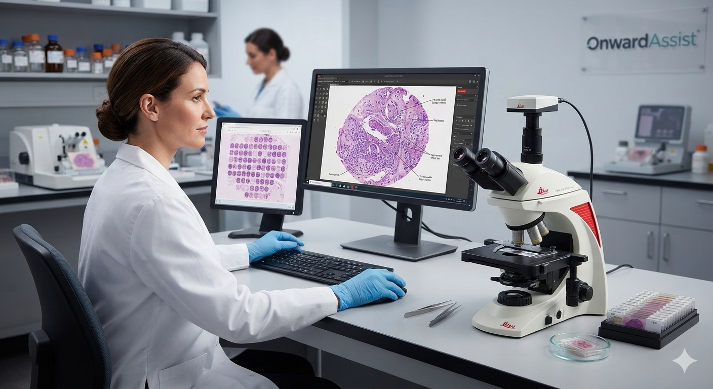
Digital pathology can support aspects of tissue management by improving access to scanned slides, facilitating review of representative tissue areas, and supporting collaboration between authorized professionals.
Depending on the system and workflow, digital slide review may help pathologists revisit a selected region, compare findings across slides, and communicate the location of relevant tissue to colleagues.
Digital records may also complement laboratory processes for documenting slide review, case progression, and relevant annotations.
However, digital images do not replace the physical tissue needed for additional sections or laboratory assays. Nor does a digital viewer, by itself, guarantee accurate tissue allocation, adequate cellularity, or successful molecular testing.
The benefits depend on integration with established laboratory procedures, accurate case identification, suitable image quality, and appropriate governance.
Digital tools should therefore be viewed as potential supports for coordination and traceability, not substitutes for sound specimen handling and diagnostic judgment.
10. Building a More Reliable Tissue Stewardship Workflow
Laboratories can strengthen tissue stewardship by making specimen adequacy, test prioritization, and communication explicit parts of their quality framework.
A practical workflow may include:
- Initial assessment: Confirm specimen identity, preservation, and the presence of representative lesional tissue.
- Diagnostic planning: Establish the morphological differential and identify the tests needed to resolve it.
- Tissue allocation: Prioritize appropriate material for essential ancillary investigations.
- Testing coordination: Align histology, immunohistochemistry, and molecular testing with assay-specific requirements.
- Adequacy review: Evaluate whether the available material is suitable for each intended test.
- Escalation: Investigate insufficient or discordant results and discuss alternatives where necessary.
- Documentation: Record relevant limitations, decisions, and outcomes according to laboratory procedures.
Quality indicators may include tissue-related test failure rates, repeat sampling attributable to specimen inadequacy, unnecessary repeat staining, molecular test success rates, and delays associated with insufficient material.
These indicators should be interpreted in the context of case mix and laboratory practice. Their purpose is to identify preventable problems and guide improvement, not to discourage clinically justified testing.
Conclusion: Preserve Tissue, Preserve Diagnostic Options
Small biopsies are an increasingly important part of modern diagnostic pathology, but their limitations require deliberate planning.
The same specimen may need to support morphological interpretation, immunohistochemistry, molecular analysis, and additional ancillary studies. Without coordinated testing and appropriate tissue stewardship, material may be exhausted before the diagnostic workup is complete.
A reliable approach combines early specimen assessment, hypothesis-driven testing, careful allocation of representative tissue, assay-specific adequacy evaluation, and clear communication between pathology and clinical teams.
The objective is not simply to conserve tissue. It is to preserve the ability to obtain the most reliable and clinically meaningful information from the specimen.
In small-biopsy pathology, diagnostic efficiency begins long before the final report. It begins with how the tissue is collected, assessed, allocated, and protected for the questions that matter most.
References and Further Reading
- Roy-Chowdhuri S, et al. Optimizing cytology and small biopsy specimen processing for ancillary studies: recommendations from the American Society of Cytopathology taskforce. Journal of the American Society of Cytopathology. 2025. Read the publication.
- Roy-Chowdhuri S, et al. Collection and Handling of Thoracic Small Biopsy and Cytology Specimens for Ancillary Studies. College of American Pathologists and collaborating professional societies. Read the guideline.
- Roh MH. The utilization of cytologic and small biopsy samples for ancillary molecular testing. Modern Pathology. 2019;32(Suppl 1):77–85. Read the publication.
Editorial note: This article is intended for professional education. Specimen handling, tissue allocation, and test selection should follow validated laboratory procedures, assay-specific requirements, current professional guidance, and case-specific clinical judgment.