



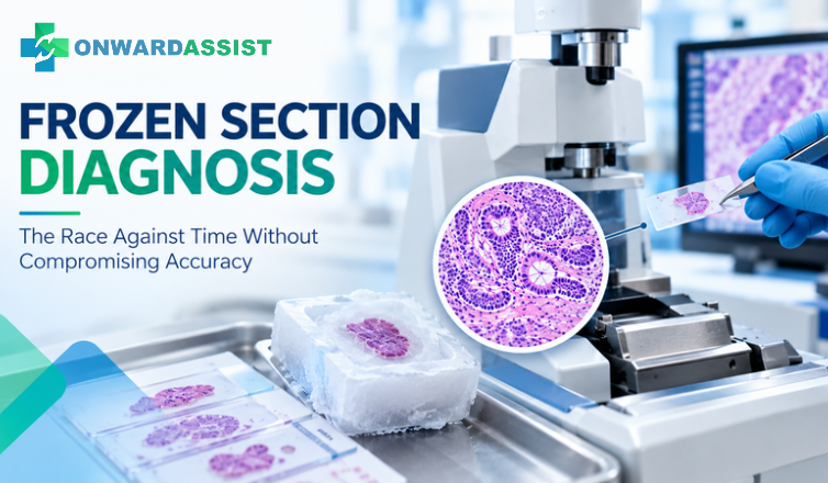
In surgical pathology, few situations demand the combination of speed, technical precision, and diagnostic judgment quite like an intraoperative frozen section examination.
During surgery, clinicians may require immediate pathological information to determine the next step in a procedure. The pathologist must assess fresh tissue, evaluate rapidly prepared frozen sections, and communicate a provisional interpretation within a limited timeframe.
The consequences of that interpretation can be significant. It may influence the extent of a surgical procedure, the decision to obtain additional tissue, the assessment of a resection margin, or whether further intervention is necessary.
However, frozen section examination is not simply a faster version of routine histopathology. Rapid freezing, restricted sampling, tissue artifacts, and limited time for evaluation introduce specific diagnostic challenges.
The central question is not how quickly a pathologist can provide an answer. It is how reliably that answer addresses the clinical question while recognizing the limitations of the material being examined.
This article explores the diagnostic challenges of frozen section examination and the practical considerations that support accurate intraoperative consultation.
1. Why Frozen Section Examination Requires a Different Diagnostic Approach
Frozen section examination is primarily used when pathological information is needed during an ongoing surgical procedure.
Common applications include:
- Assessing surgical resection margins.
- Evaluating whether a suspicious lesion represents malignancy.
- Identifying tissue submitted for confirmation, such as lymph node or parathyroid tissue.
- Assessing selected lesions when the result may influence immediate surgical management.
- Determining whether additional tissue sampling is required.
Unlike routine paraffin histology, frozen section examination operates within a compressed diagnostic window.
Fresh tissue is received, appropriately sampled, embedded in freezing medium, rapidly frozen, sectioned using a cryostat, stained, and examined microscopically.
The result is then communicated to the surgical team.
This process requires close coordination between the operating room, specimen reception, histotechnology personnel, and the pathologist.
An important consideration is whether the requested examination will actually influence immediate patient management. If the result will not change an intraoperative or perioperative decision, routine or expedited permanent-section examination may be more appropriate.
Frozen section is therefore most valuable when there is a clearly defined clinical question that can reasonably be answered using the available tissue and technique.
2. Freezing Artifacts: When Tissue Processing Changes the Morphology
One of the most important limitations of frozen section examination is the introduction of technical artifacts during rapid tissue freezing and section preparation.
In routine histopathology, tissue undergoes controlled fixation and processing before paraffin embedding. Frozen sections follow a different preparation pathway, which can compromise morphological detail.
Common technical difficulties include:
Ice crystal artifacts
Rapid freezing can produce ice crystal formation, resulting in spaces or distortions within tissue. These changes may interfere with the assessment of cellular detail and tissue architecture.
Compression and folding artifacts
Mechanical compression, section folding, and uneven section thickness can obscure cellular relationships and create misleading appearances.
Poor section quality
Difficult tissue consistency, inappropriate freezing conditions, and technical variation during cryostat sectioning can affect the interpretability of the slide.
Tissue fragmentation
Fragile or poorly preserved tissue may fragment during preparation, making it difficult to evaluate the relationship between different tissue components.
These artifacts are not merely technical inconveniences. They can directly influence the pathologist’s ability to distinguish reactive changes, atypia, and malignancy.
Recognizing when morphology has been compromised is essential. A technically suboptimal slide should not be interpreted with the same confidence as a well-preserved permanent section.
3. Sampling Error: The Diagnostic Problem Beyond the Microscope
A frozen section interpretation can be technically accurate for the tissue examined and still fail to represent the overall lesion.
This is the fundamental challenge of sampling error.
A specimen may contain heterogeneous areas with different morphological characteristics. A small selected fragment may not include the area that ultimately determines the final diagnosis.
This becomes particularly important in:
- Heterogeneous neoplasms.
- Lesions with focal invasion.
- Small suspicious areas within otherwise benign tissue.
- Tumours with extensive necrosis or fibrosis.
- Complex surgical margins.
- Specimens in which the lesion is difficult to localize grossly.
For example, a lesion may contain predominantly benign-appearing tissue with a small focus of invasive disease. If the selected frozen tissue does not include that focus, the intraoperative assessment may differ from the final permanent-section diagnosis.
The same issue can arise during margin assessment. A negative frozen section reflects the tissue and levels actually examined, not necessarily every microscopic area of the entire surgical margin.
Careful gross examination, orientation, and selection of representative tissue are therefore fundamental components of frozen section accuracy.
The pathologist should also consider whether the requested question can be answered adequately from the submitted material.
4. Tumour Heterogeneity and the Difficulty of Recognizing Invasion
Tumour heterogeneity introduces additional complexity into intraoperative interpretation.
Different regions of a lesion may demonstrate different degrees of differentiation, architectural patterns, stromal relationships, or invasive characteristics.
In permanent sections, additional levels and broader sampling may help establish these relationships. Frozen section offers fewer opportunities for extensive examination.
Certain diagnostic distinctions are particularly difficult in frozen tissue.
Examples include:
- Reactive epithelial atypia versus malignancy.
- Fibrosis associated with inflammation versus desmoplastic stromal response.
- Benign glandular structures versus invasive glandular growth.
- Mesothelial cells versus metastatic carcinoma.
- Well-differentiated neoplastic tissue versus non-neoplastic mimics.
These distinctions may depend on subtle cytological and architectural features that are less reliably preserved in frozen sections.
The presence of an atypical structure should therefore be interpreted in the context of its surroundings, the clinical history, and the specific surgical question.
When the defining features of invasion or malignancy cannot be established confidently, a definitive intraoperative classification may not be justified.
5. Margin Assessment: What Does a Negative Frozen Section Actually Establish?
Intraoperative margin assessment is one of the important applications of frozen section examination.
Surgeons may request evaluation of a specific margin to determine whether additional tissue should be removed.
However, margin interpretation requires careful consideration of specimen orientation, tissue selection, and the relationship between the sampled section and the actual surgical margin.
Several factors can complicate the assessment:
Incorrect or incomplete orientation
If the anatomical orientation is unclear, the pathologist may be unable to establish which surface or region corresponds to the requested margin.
Inadequate tissue representation
The submitted frozen section may not include the area of greatest concern.
Tissue distortion
Freezing, compression, cautery, and other artifacts can interfere with the assessment of the tumour-to-margin relationship.
Complex anatomical surfaces
Some specimens contain irregular or multiple margins that require careful identification and mapping.
A negative frozen section should therefore be interpreted within the boundaries of the sampled tissue and the method used.
Clear communication is important. The report should specify the evaluated margin and avoid implying that the entire specimen has been examined when only selected areas were assessed.
6. When Should a Pathologist Defer a Frozen Section Diagnosis?
One of the most important aspects of intraoperative consultation is recognizing when an immediate definitive answer is not justified.
Deferral is not a diagnostic failure. It can be the most appropriate decision when technical limitations or morphological uncertainty prevent a reliable conclusion.
Situations that may warrant deferral include:
- Severe freezing artifact.
- Insufficient or non-representative tissue.
- A lesion requiring extensive architectural assessment.
- Borderline or equivocal morphological findings.
- A differential diagnosis requiring immunohistochemistry or molecular studies.
- A distinction that cannot be reliably established using frozen tissue.
- A result that requires additional sampling or permanent-section evaluation.
A deferred diagnosis should communicate what has been observed, why a definitive interpretation cannot be provided, and what further evaluation is required.
The pathologist should also consider whether additional tissue can be safely obtained or whether the surgeon’s immediate question can be answered in another way.
The key is to distinguish between three different situations:
- A definitive interpretation is supported by the available tissue.
- The findings are suspicious or limited but do not support a definitive classification.
- The examination cannot resolve the clinical question and requires permanent-section evaluation.
These distinctions help prevent uncertainty from being communicated as certainty.
7. The Communication Gap Between the Pathologist and Surgeon
Frozen section diagnosis does not end when the pathologist reaches an interpretation.
The result must be communicated accurately to the operating surgeon, often within a short period and under time-sensitive conditions.
Even when the pathological interpretation is correct, unclear communication can lead to misunderstanding.
Consider the difference between:
- No malignancy identified in the examined frozen sections.
- Findings are suspicious for malignancy.
- The examination is deferred pending permanent sections.
These statements communicate different levels of certainty and have different implications for surgical decision-making.
Terms such as “favor,” “suspicious,” or “cannot exclude” should be used carefully, with sufficient context to explain the diagnostic limitation.
A reliable communication process should include:
- Confirmation of patient and specimen identification.
- Confirmation of the specific clinical question.
- Clear identification of the specimen or margin being evaluated.
- Direct communication of the result to the appropriate surgical professional.
- Explicit explanation of deferred or uncertain findings.
- Documentation of the communication according to institutional procedures.
Where appropriate, a read-back process can help confirm that the surgeon has understood the result correctly.
Effective intraoperative consultation is a shared process. Diagnostic accuracy depends not only on the interpretation but also on the clarity with which the information reaches the clinical team.
8. Frozen Section Discordance: Why Permanent Sections Remain Essential
Frozen section interpretation is provisional and must be correlated with the final permanent-section examination.
Discordance may arise from several different sources:
Sampling-related discordance: The relevant lesion or diagnostic feature was not represented in the frozen tissue.
Technical discordance: Freezing artifacts or section quality obscured important morphological details.
Interpretive discordance: The available findings were misclassified or an important differential diagnosis was not adequately considered.
Communication-related discordance: The original interpretation was not accurately conveyed or understood.
A 2023 study published in the American Journal of Clinical Pathology evaluated 6,910 frozen section cases and reported an overall discordance rate of 2.9%. The study also highlighted the importance of subspecialty familiarity and access to prior material in reducing major interpretive discrepancies.
This illustrates why frozen section performance should not be evaluated solely through a single overall accuracy figure.
Laboratories should review the nature and clinical significance of discrepancies, identify their underlying causes, and use the findings to improve practice.
The final permanent-section diagnosis remains essential for establishing the definitive pathological interpretation.
9. Quality Assurance: Measuring More Than Turnaround Time
Frozen section performance is often associated with the time required to deliver a result. However, turnaround time alone does not adequately measure diagnostic quality.
A laboratory may produce slides rapidly but still experience problems with specimen orientation, sampling, technical preparation, interpretation, or communication.
A more complete quality framework should evaluate different stages of the process.
Pre-analytical quality
- Correct specimen identification.
- Adequate clinical information.
- Clear communication of the surgical question.
- Appropriate specimen transport and handling.
Analytical quality
- Representative tissue selection.
- Appropriate freezing conditions.
- Acceptable section quality.
- Reliable microscopic interpretation.
Post-analytical quality
- Timely and accurate communication.
- Clear documentation.
- Appropriate handling of deferred diagnoses.
- Correlation with permanent sections.
Useful quality indicators may include frozen-to-permanent-section discordance, deferral rates, major diagnostic discrepancies, specimen-related delays, technical repeat rates, and communication-related incidents.
Importantly, a higher deferral rate does not automatically indicate poor performance. Appropriate deferral may reflect responsible recognition of diagnostic limitations.
Quality assessment should examine whether the pathologist made an appropriate decision based on the available evidence, rather than rewarding definitive answers in every case.
10. The Role of Digital Pathology in Intraoperative Consultation
Digital pathology introduces additional opportunities for image access, consultation, and collaboration in surgical pathology.
In settings where validated digital systems are available, scanned images can support remote review and facilitate communication between pathologists at different locations.
This may be particularly relevant to institutions with multiple laboratories, distributed pathology teams, or limited access to particular subspecialty expertise.
Potential applications include:
- Remote review of selected histological images.
- Consultation between pathologists.
- Access to prior digital slides where available.
- Documentation and sharing of relevant microscopic findings.
- Support for multi-site pathology workflows.
However, digital review does not automatically eliminate the technical limitations of frozen tissue.
Image quality, scanning time, network performance, display conditions, system validation, and the intended clinical use all require careful evaluation.
A digital image also represents only the tissue that has actually been sampled and prepared. It cannot correct an inadequate specimen selection or restore morphological detail lost during freezing.
For this reason, digital pathology should complement established intraoperative consultation procedures rather than be viewed as a substitute for appropriate specimen handling and professional diagnostic judgment.
Conclusion: Speed Matters, but Diagnostic Judgment Matters More
Frozen section examination remains an important part of surgical pathology because it provides timely information when immediate clinical decisions depend on pathological assessment.
Its value, however, depends on recognizing its limitations.
Sampling error, freezing artifacts, tumour heterogeneity, margin orientation, interpretive uncertainty, and communication challenges can all influence the final outcome.
Reliable intraoperative consultation requires more than rapid slide preparation. It requires careful specimen selection, clear clinical questions, disciplined morphological interpretation, appropriate use of deferral, and effective communication with the surgical team.
The objective is not to provide a definitive answer in every situation. It is to provide the most reliable information that the available tissue and examination method can support.
In frozen section diagnosis, the most valuable decision is not always the fastest diagnosis. Sometimes, it is knowing precisely what the tissue can—and cannot—tell us.
References and Further Reading
- Patterns of Major Frozen Section Interpretation Error: An In-Depth Analysis From a Complex Academic Surgical Pathology Practice. American Journal of Clinical Pathology. 2023.
https://pubmed.ncbi.nlm.nih.gov/37141256/ - Quantitative and Qualitative Assessment of Frozen Section Diagnosis Accuracy and Deferral Rate Across Organ Systems. American Journal of Clinical Pathology.
https://doi.org/10.1093/ajcp/aqac115 - College of American Pathologists and National Society for Histotechnology. Practical Guide to Specimen Handling in Surgical Pathology. Version 11.0, September 2023.
https://documents.cap.org/documents/practical-guide-specimen-handling.pdf - Royal College of Pathologists of Australasia. Frozen Section – RCPA Manual.
https://www.rcpa.edu.au/Manuals/RCPA-Manual/Pathology-Tests/F/Frozen-section - American Society for Clinical Pathology. Do Not Order a Frozen Section on a Pathology Specimen if the Result Will Not Affect Immediate Patient Management.
https://www.ascp.org/news/effective-test-utilization/effective-test-utilization-recommendations/effective-test-utilization-recommendation-details/5710aefb-922c-492c-98ee-95c56e47fe31
Editorial note: This article is intended for professional education and discussion. Frozen section indications, specimen handling, reporting, and quality requirements should follow institutional protocols, current professional guidance, and case-specific clinical judgment.