



How Deep Learning Is Advancing Cell Detection, Mitotic Counting and Histological Grading
Introduction
In histopathology, some of the most important diagnostic information is contained in individual cells.
Mitotic figures are among these important features. They represent cells undergoing division and provide an indication of proliferative activity within a tissue.
In several tumour grading systems, mitotic activity contributes to the assessment of biological behaviour and can influence the final histological grade.
However, identifying and counting mitotic figures is not always straightforward.
Pathologists must distinguish genuine mitotic figures from apoptotic bodies, pyknotic nuclei, inflammatory cells and other structures that may appear morphologically similar. They must also identify appropriate regions for counting, assess cellular morphology and apply the relevant grading criteria.
This process can be time-consuming, particularly when examining large tissue sections.
With the development of digital pathology and deep learning, researchers are investigating whether artificial intelligence can help identify mitotic figures, quantify their distribution and support more consistent proliferation assessment.
Recent studies published in 2025 and 2026 demonstrate meaningful progress in this area.
The important question is no longer simply whether AI can recognize a dividing cell. It is whether automated mitotic analysis can become sufficiently reliable, reproducible and clinically useful across different tissue types and laboratory environments.
1. Why Mitotic Figure Assessment Matters
Mitotic activity reflects the rate at which cells are undergoing division.
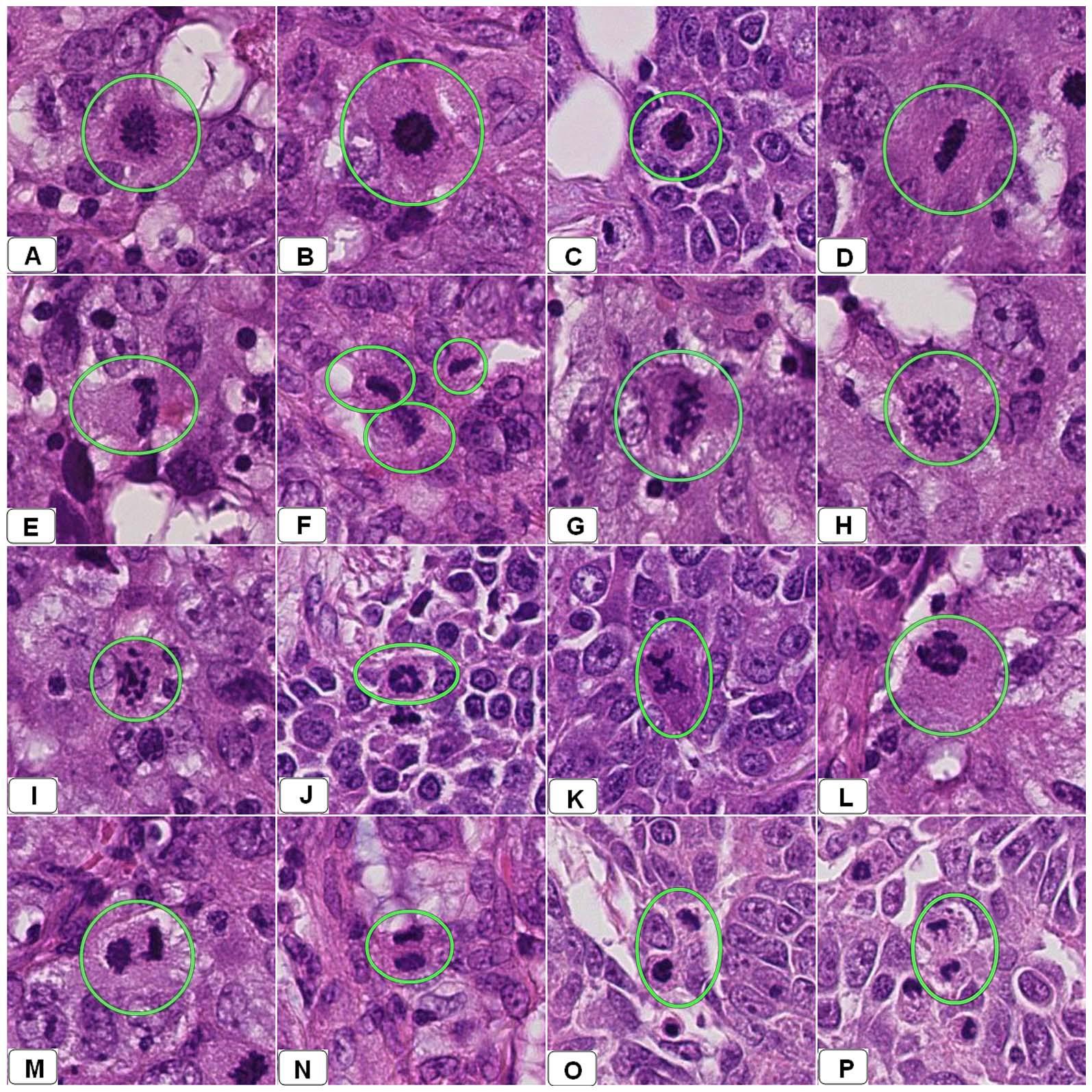
In histopathological examination, mitotic figures are identified through characteristic changes in nuclear morphology during cell division.
Depending on the tumour type and grading system, mitotic activity may contribute to:
- Histological grading
- Assessment of cellular proliferation
- Prognostic evaluation
- Research into tumour behaviour
- Comparison of tissue characteristics across cases
For example, mitotic count is one of the components of the Nottingham grading system used in breast carcinoma assessment.
In other tumour types, mitotic activity may be considered alongside additional morphological features, including cellular atypia, tissue architecture and necrosis.
However, the significance of a mitotic count is not universal. It depends on the tumour type, the applicable grading system, the area examined and the method used to quantify mitotic activity.
This makes accurate identification and appropriate interpretation particularly important.
2. Why Manual Mitotic Counting Can Be Challenging
Although mitotic counting is an established histopathological task, it involves several practical difficulties.
A. Distinguishing mitotic figures from mimickers
Not every dark or condensed nucleus represents a mitotic figure.
Apoptotic bodies, pyknotic nuclei, crushed cells, inflammatory cells and other structures can resemble mitotic figures.
Accurate identification requires careful assessment of nuclear morphology and the relevant stage of mitosis.
B. Selecting the appropriate counting region
Mitotic activity may not be evenly distributed throughout a tissue section.
Some regions may demonstrate higher proliferative activity than others.
The selection of representative or mitotically active areas can therefore influence the final count.
C. Examining large tissue sections
With whole-slide imaging, a single tissue section can contain millions of cells.
Identifying individual mitotic figures across large digital images requires considerable visual attention, particularly when the relevant structures are relatively small.
D. Counting and recording
Manual counting requires the pathologist to identify each qualifying figure while avoiding omissions and duplicate counts.
This becomes especially challenging when several regions must be assessed or when multiple slides are involved.
E. Differences in interpretation
Morphologically ambiguous figures can be interpreted differently by different observers.
The challenge is not simply counting cells. It is consistently determining which structures meet the criteria for a mitotic figure.
These difficulties have encouraged researchers to investigate automated detection and quantification approaches.
3. How Deep Learning Detects Mitotic Figures
AI-based mitotic figure detection generally involves several computational stages.
A model is trained using histopathology images in which mitotic figures have been identified and annotated by expert reviewers.
It learns visual characteristics that help distinguish mitotic figures from surrounding tissue structures.
Depending on the architecture, the system may use object detection, image classification or segmentation.
A typical computational pipeline includes the following stages.
Stage 1: Tissue identification
The digital slide is examined to identify tissue-containing regions and exclude irrelevant background areas.
This helps reduce unnecessary image processing.
Stage 2: Candidate detection
The model scans selected image regions and identifies structures that may represent mitotic figures.
At this stage, the system aims to identify potential candidates rather than make a definitive diagnostic interpretation.
Stage 3: Candidate classification
Detected structures are examined more closely to distinguish genuine mitotic figures from visually similar non-mitotic structures.
A second-stage classifier can help reduce false-positive detections.
Stage 4: Localization and quantification
The system records the location of detected figures and can calculate counts within defined tissue regions.
Stage 5: Expert review
A pathologist reviews the findings, assesses uncertain candidates and determines whether the results are appropriate for the intended clinical or research application.
This combination of automated detection and professional review is important because a model’s output depends on its training data, image quality and intended use.
4. Recent Research: What Is Changing in AI-Based Mitosis Detection?
Several recent studies provide useful insight into the progress of automated mitotic analysis.
A. Pan-Cohort Validation: Can Deep Learning Count Mitoses Across Different Tumour Types?
A significant study published in FEBS Open Bio on 12 February 2026 investigated the applicability of deep-learning-based mitotic figure counting across multiple tumour types.
The researchers developed a model using H&E-stained whole-slide images from a publicly available breast cancer dataset containing expert-annotated mitotic figures.
The final model was externally evaluated using 14,571 patient samples from 13 patient cohorts representing seven different tumour types.
The researchers examined the relationship between automatically detected mitotic figures per square millimetre and clinical outcomes.
Automated mitotic counts correlated with known proliferation rates. Higher mitotic counts were associated with significantly worse patient outcomes in the studied tumour types, with colorectal cancer being the exception.
Why this research matters:
The study moves beyond evaluating a model on a single image dataset. Its multi-cohort validation provides important evidence about the potential applicability of automated mitotic counting across different tumour settings.
However, the findings do not establish that one automated counting approach can replace tumour-specific grading criteria or professional interpretation.
Research paper:
B. A Three-Stage AI Pipeline for Mitotic Detection and Classification
A study published in Cancers on 1 June 2026 investigated a scalable deep-learning pipeline for mitotic detection and analysis.
The researchers developed a three-stage approach:
- A YOLOv11-based detector to identify candidate mitotic figures.
- A lightweight classifier to refine detections and suppress false positives.
- A downstream classifier to distinguish atypical from normal mitotic figures.
The study evaluated the pipeline using public mitosis detection and atypical mitosis classification benchmarks. It was also applied to breast cancer whole-slide images from the TCGA-BRCA cohort.
The researchers reported that the pipeline could process gigapixel whole-slide images within minutes on a single GPU.
An exploratory survival analysis suggested that mitosis-derived features might provide modest additional prognostic information beyond clinical and nuclear morphology features.
Why this research matters:
This work demonstrates that automated mitotic analysis is evolving beyond simply detecting potential dividing cells.
The classification of atypical mitotic figures introduces another level of morphological assessment that may be relevant to research into cellular behaviour.
The reported findings remain research evidence and require further independent clinical validation.
Research paper:
C. Active Learning: Reducing the Need for Extensive Manual Annotation
One of the major challenges in developing pathology AI models is the need for accurately annotated training data.
Identifying mitotic figures requires expert review, and preparing large annotated datasets can be resource-intensive.
A study published in Histopathology in October 2025 investigated a deep active-learning framework for mitotic figure detection in glioblastoma whole-slide images.
Instead of relying exclusively on a large, fully annotated dataset, the framework initially trained a convolutional neural network using a smaller annotated dataset.
It then identified uncertain examples from an unlabelled data pool. These selected examples were reviewed by experts and incorporated into subsequent model training.
The study reported:
- 81.75% precision for mitotic figure detection.
- 82.48% recall for mitotic figure detection.
- 84.1% accuracy for mitotic figure subclass classification.
- Approximately 900 minutes of annotation time across 66 whole-slide images, reducing the annotation effort by nearly half compared with the traditional approach described in the study.
Why this research matters:
Active learning offers a practical strategy for developing pathology AI systems when expert annotation is expensive.
Rather than asking pathologists to label every available image, the system prioritizes examples where additional expert input may be most useful.
Research paper:
D. MIDOG++: Addressing the Problem of Different Scanners and Tissue Types
A major challenge in computational pathology is that images acquired in different laboratories may not look identical.
Staining intensity, tissue preparation, scanner characteristics and image acquisition settings can all affect how tissue structures appear.
These variations can influence the performance of AI models trained on images from a limited number of sources.
The MIDOG++ dataset, published in Scientific Data in 2023, was developed to support research into this problem.
It contains 503 histological specimens across seven tumour types and 11,937 annotated mitotic figures.
The dataset incorporates variation across laboratories, tissue types and five whole-slide scanners.
The researchers demonstrated that domain shifts can affect mitotic detection performance and investigated approaches to improve generalization.
Why this research matters:
A mitotic detection model should not be evaluated only on images that closely resemble its training data.
Testing across scanners, tissue types and laboratory conditions is essential when assessing whether a system may be useful beyond a controlled research environment.
Research paper:
A Comprehensive Multi-Domain Dataset for Mitotic Figure Detection — Scientific Data (2023)
5. What Makes Mitotic Figure Detection Difficult for AI?
Despite encouraging research results, automated mitotic detection remains a technically demanding task.
1. Morphological similarity
Mitotic figures can resemble several non-mitotic structures.
A model must learn subtle morphological differences rather than simply identify dark or condensed nuclei.
2. Class imbalance
Mitotic figures are relatively uncommon compared with the large number of other cellular structures in a tissue section.
This creates an imbalanced detection problem in which false positives and missed detections can both affect performance.
3. Variation in mitotic morphology
Different stages of mitosis have different appearances.
A model trained on a narrow range of morphological examples may struggle with less common or atypical forms.
4. Tissue and staining variation
Differences in fixation, staining, section thickness and scanner characteristics can affect image appearance.
A model that performs well on one dataset may not maintain the same performance across different laboratories.
5. Region selection and spatial distribution
Mitotic activity is not always uniform throughout a tissue section.
An automated system must be evaluated not only for its ability to recognize individual figures, but also for how it handles the selection and quantification of relevant regions.
6. False-positive and false-negative detections
False positives can inflate a mitotic count, while false negatives can lead to underestimation.
Both errors may have consequences when counts are used in a grading workflow.
This is why sensitivity, precision, recall, localization accuracy and performance across independent datasets should all be considered.
6. Beyond Counting: What Could Automated Mitotic Analysis Support?
The potential value of AI-based mitotic detection extends beyond generating a numerical count.
When appropriately validated, automated analysis may support several research and workflow applications.
Quantitative assessment
AI can identify and localize candidate mitotic figures, helping researchers investigate cellular proliferation across defined tissue regions.
Atypical mitosis analysis
Models may help classify different morphological categories of mitotic figures, supporting research into atypical cellular division.
Large-scale retrospective studies
Automated analysis can make it more practical to investigate mitotic activity across large digital slide collections.
Reproducibility research
AI-assisted counting provides an opportunity to study how automated measurements compare with expert assessments and established grading methods.
Research into prognostic features
Quantitative mitotic features may be investigated alongside other histological and clinical variables to explore associations with patient outcomes.
These applications should be considered in the context of their validation evidence and intended use.
7. What Should Be Evaluated Before Clinical Implementation?
A promising model is not automatically a clinically suitable model.
Before integrating automated mitotic detection into a diagnostic environment, several factors require consideration.
- Analytical performance: How accurately does the model detect and localize mitotic figures?
- External validation: Does performance remain acceptable on independent datasets?
- Scanner compatibility: Has the model been tested using images from different scanner manufacturers?
- Staining variation: How does it respond to differences in staining intensity and tissue preparation?
- Tumour-specific applicability: Is the model validated for the intended tumour type and grading system?
- Workflow integration: Can the results be reviewed and interpreted efficiently by pathologists?
- Traceability: Can detected candidates, reviewed regions and analysis outputs be documented?
- Clinical governance: Are the model’s intended use, limitations and professional responsibilities clearly defined?
A particularly important consideration is the distinction between detecting mitotic figures and assigning a histological grade.
A mitotic count is only one component of the relevant grading system in many settings. The final interpretation must account for all applicable diagnostic criteria.
AI-generated counts should therefore be treated as supporting information unless the specific application has been appropriately validated and authorized.
8. The Role of Digital Pathology Infrastructure
The development of mitotic detection models also highlights the importance of the digital pathology environment in which AI analysis takes place.
A model may be capable of identifying thousands of candidate structures, but its practical value depends on how those results are managed and reviewed.
A connected digital pathology platform should support:
- High-resolution whole-slide image access.
- Efficient navigation between image regions.
- Annotation and region-of-interest management.
- Integration of suitable AI analysis tools.
- Structured case and reporting workflows.
- Review of AI-generated findings.
- Secure access and role-based permissions.
- Traceability of analysis and review activities.
These capabilities help connect computational analysis with the broader pathology workflow.
At Onward Assist, our focus is on building connected digital pathology workflows that bring together image management, case coordination, reporting and AI-assisted capabilities.
As research advances in areas such as mitotic detection, the ability to integrate analytical tools into organized and traceable digital environments will become increasingly relevant.
Conclusion: From Manual Counting to AI-Assisted Quantification
Mitotic figure assessment is a well-established component of histopathological practice, but identifying and quantifying mitotic activity can be demanding.
Recent developments in deep learning demonstrate how AI can support candidate detection, classification, quantification and large-scale research analysis.
The progression from conventional image classification to multi-stage detection pipelines, active learning and external multi-cohort validation is particularly encouraging.
However, reliable clinical implementation requires more than promising detection metrics. Models must demonstrate performance across relevant tissue types, laboratories, scanners and clinical settings.
The objective should not be to replace the pathologist’s assessment, but to investigate whether automated tools can provide useful, reproducible and reviewable quantitative support.
The future of AI-assisted mitotic assessment will depend not only on how accurately a model detects a dividing cell, but also on how reliably that detection contributes to the overall histopathological interpretation.
References and Further Reading
- Applicability of mitotic figure counting by deep learning: a development and pan-cancer validation study. FEBS Open Bio. 2026.
https://doi.org/10.1002/2211-5463.70210 - Scalable and Efficient Deep Learning-Based Pipeline for Mitotic Detection and Analysis in Pathology Images. Cancers. 2026.
https://doi.org/10.3390/cancers18111807 - Liu E, et al. A deep active learning framework for mitotic figure detection with minimal manual annotation and labelling. Histopathology. 2025.
https://doi.org/10.1111/his.15506 - A comprehensive multi-domain dataset for mitotic figure detection. Scientific Data. 2023.
https://www.nature.com/articles/s41597-023-02327-4 - A deep learning framework deploying segment anything to detect pan-cancer mitotic figures from haematoxylin and eosin-stained slides. Communications Biology. 2024.
https://www.nature.com/articles/s42003-024-07398-6 - Mitosis detection, fast and slow: Robust and efficient detection of mitotic figures. Medical Image Analysis. 2024.
https://doi.org/10.1016/j.media.2024.103052
Editorial note: This article discusses research into AI-assisted mitotic figure detection and quantification. Reported model performance is specific to the datasets and methods used in each study and should not be interpreted as evidence of universal clinical performance. Clinical use requires appropriate validation, regulatory assessment where applicable, institutional governance and pathologist oversight.